
Scientific photography is a type of photography that will impress you by The images that are usually captured since they are usually very unusual, if you like the world of science and discovery, this type of photography may be for you, that is why in this article you will find the necessary information to know and discover if the Scientific photography may be your thing.
Background of scientific photography
Science and photography have been intertwined to a great extent since the invention of photography, even though photography has always been associated with aesthetic or documentary aspects. Photography has been used and used by scientists for many everyday purposes. Within the history of photography are several events that contribute to the training of scientific photography,
During the beginning of microscopes in the 17th century, microscopists have had a need to describe their observations. The first photomicrographs were by Thomas Wedgwood and Humphrey David in 1802 a solar microscope was used, unfortunately these were images of short duration because a permanent processing method for photographic images had not yet been developed.
W. Talbot was the first photographer interested in microscopic optical techniques , in 1837, using a solar microscope he managed to obtain optical drawings of crystals in polarized light, micrographs of an insect wing were shown.
What is scientific photography?
Science is a system of verifiable knowledge therefore scientific studies require having a support or a record, which is why the objective of the Scientific photography is to illustrate and fix that record, on the other hand, scientific photography facilitates the observation of different phenomena that cannot be easily seen if not through a lens.
Scientific photography is a set of photographic knowledge applied in scientific documentation, recording and observations. In scientific photography it is possible to take photographs with lengths wavelength from 10 to 1200 nanometers, using special cameras combined with projectors and filters to achieve the desired wavelength. This is useful in fields such as phytopathology research, agriculture, astronomy, medicine, etc.
There are specialized cameras for each branch of scientific photography since situations and needs can be very diverse, from very slow shots or vice versa to operations extremely fast. An example of a second approach is high-speed imaging, which can take more than 55,000 images per second, with which it is possible to reconstruct and control the true movement of the human sperm tail.
Importance of scientific photography
We can understand the importance of scientific photographs by comparing the limited vision of the human eye with the phenomena of nature, which is why this tool is very useful to be able to capture the imperceptible to the human eye.
Optical instruments such as microscopes, telescopes, or spectrometers are the instruments used to capture scientific images. Thanks to this, much progress has been made in science. In 1953 the image representing the double helix structure of DNA was captured and obtained using X-ray diffraction techniques.

Types of scientific photography
Macro
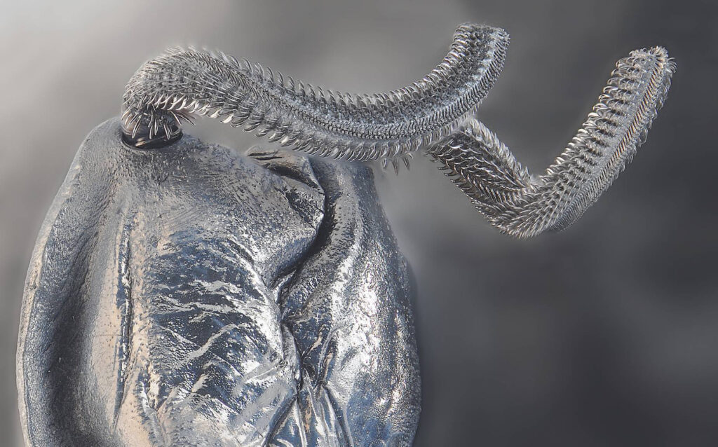
Photomicrography or macrophotography are images of the small world, that is, plants, animals. Normally it is used for very small objects, so these images are larger or equal in size to the photographed subject.
The use of this type of photography is frequently used in scientific photography within biological research, since with it it is possible to document and study species of the animal world as a vegetable.

Micro
For smaller species scientific photography uses microphotography, these photographs are captured with different types of optical microscopes. The difference between photomicrography and photomicrography is that it resorts to the use of a microscope and can obtain photographs with a magnification of 10X onwards, which allows images to be taken.
Scientific micro photography captures extremely small images, difficult to see, the image is obtained through a microscope and photographed with a camera adapted to a microscope, for development Microscopes are designed to incorporate a photographic camera, however it is possible that you can add a camera to any microscope since there are various external attachments for this.

Ultraviolet
Ultraviolet photography is a process of obtaining images that are recorded using only ultraviolet light. Ultraviolet images serve a variety of scientific, medical or artistic purposes, they can reveal the deterioration of works of art or structures that are not visible under visible light.
Diagnostic medical images can be used to detect specific skin disorders or as evidence of infection. Some animals, especially insects, use ultraviolet wavelengths to see; Also ultraviolet images can help to study the markings on plants that attract insects, when they are not visible to the naked eye, in archaeological sites UV images can reveal artifacts or traffic patterns that would otherwise be invisible.
In ultraviolet photography uses a lamp that emits this type of ultraviolet light to illuminate a subject. By placing a special filter in front of the viewfinder, visible light is blocked and ultraviolet rays are allowed to pass through. The filter is made of specially tinted glass. Photographers find UV filters to be an effective way to block visible light, allowing only lenses to be illuminated by UV rays.
